


4 Things to Check During a Demo Before Buying a Skin Analysis Device
When choosing a skin analysis device for a dermatology practice, it is difficult to judge its actual performance from specifications and sample images alone. Before purchasing, it is helpful to use a demo to check image quality, lighting quality, consistency across repeated captures, analysis results, and progress comparison features firsthand.
These elements provide the foundation for observing patients’ skin in detail, interpreting image-based analysis results, and assessing changes before and after treatment. This article introduces four things dermatologists should check during a skin analysis device demo.
1. Does repeated imaging produce images that can be reliably compared?
Consistent imaging conditions are essential for monitoring changes in the skin. Differences in lighting, facial angle, camera distance, or expression can make skin tone and wrinkles appear different even when the skin itself has not changed. This is why standardization matters in clinical photography.
During the demo, photograph the same person once, then ask them to move their face away from the device, reposition themselves, and take another image. Check how consistently the device reproduces facial position and size, skin color, and brightness in key areas.
It is also helpful to ask about facial positioning guidance, lighting and exposure control, and calibration methods used to maintain consistent color and brightness. If analysis values differ between repeated captures, request repeatability data and the test conditions for the relevant measurement.
Although a single demo cannot fully verify repeatability, it can show how easily users can reproduce the required imaging conditions.
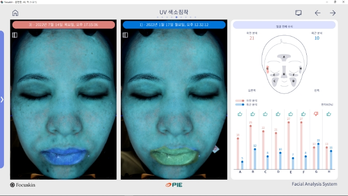
2. Does each imaging mode clearly reveal information relevant to clinical practice?
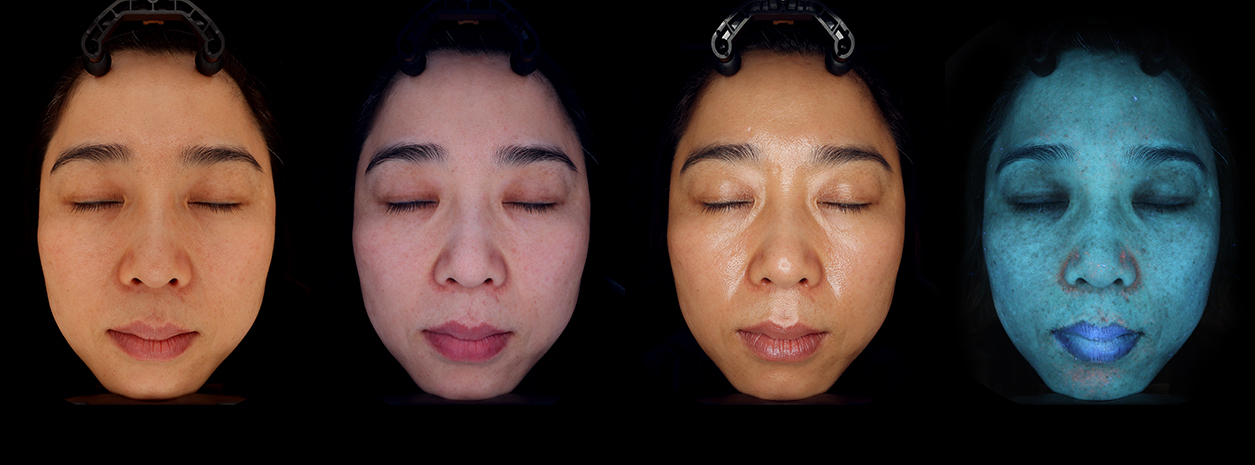
Standard-light, polarized-light, and UV imaging allow the skin to be observed under different optical conditions. More important than the number of modes is whether they provide images suited to the skin features you want to observe.
Light reflected from the skin includes both surface reflections and light that returns after scattering within skin tissue. Polarized imaging makes use of these properties. Parallel polarization emphasizes surface detail, while cross-polarization reduces surface reflections and helps reveal pigmentation and vascular-related coloration.
For example, cross-polarized images are useful for examining redness and pigment distribution, but standard-light images should also be reviewed when assessing surface morphology. Because cross-polarization reduces reflections, skin texture and the degree of lesion elevation may be less apparent.
During the demo, switch between imaging modes for the same area and check which features become clearer. For UV mode, review actual captured images and ask for an explanation of the device’s imaging method and analysis parameters.
3. Are image quality and lighting quality sufficient to capture fine skin details?
In a skin analysis device, captured images serve both as visual records for clinical observation and as input data for analysis. Image quality and lighting quality are therefore essential factors to check when choosing a device.
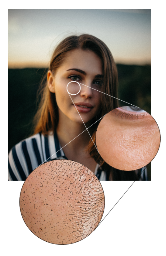
Pixel-based image analysis uses information such as brightness, color, boundaries, and patterns recorded in an image. If focus is poor or detail is insufficient, the boundaries of small pores and fine wrinkles may not be clearly captured, while excessive reflections or shadows can obscure the features being analyzed. Alongside the performance of the analysis algorithm, check how faithfully the underlying images capture skin features.
During the demo, look beyond the full-face image and zoom in on areas such as the cheeks, eye area, and forehead. Check whether pore boundaries and fine wrinkles are distinguishable, whether bright areas are washed out, and whether details are lost in dark areas. In addition to the camera’s pixel count, check the maximum image magnification, lens, focus, exposure, and lighting.
Lighting determines how skin features appear in an image. Its direction and conditions are particularly important when recording subtle surface features and skin texture. In standard-light imaging, check whether skin color appears natural and consistent, and whether reflections or shadows interfere with observation in any facial area. In polarized-light and UV imaging, check whether the features relevant to each mode’s purpose are clearly visible.
Good imaging quality provides the foundation for detailed clinical observation and for capturing the information required for image analysis. During the demo, check both the clarity of the display and whether the images actually contain the fine details needed for analysis.
4. Does the device fit your clinical workflow, and is support available when problems arise?
Even a device with extensive features can be difficult to use consistently in a clinical setting if image capture and result review are complicated. During the demo, experience the entire process, from patient registration to imaging, analysis, record storage, and explanation of the results.
In particular, if staff capture the images and a dermatologist reviews the results, it is important that different operators can follow the same procedure. Also check whether patient records are easy to find, required images can be opened quickly, and results can be printed or shared.
Post-purchase support is also part of choosing a device. Ask whether there is a clear point of contact when problems arise, whether support staff can review the screen remotely and provide guidance, and what procedures apply when an on-site inspection or action involving parts is required.
PIE Co., Ltd. responds promptly and courteously to usability issues and software problems, helping resolve them by reviewing the operating screen and settings remotely. Practical support is available, from guidance on device operation to troubleshooting.
A skin analysis device demo should include repeated imaging, switching between imaging modes, reviewing the areas being analyzed, comparing changes over time, and checking day-to-day operation and support.
During a demo of PIE’s JANUS Pro or Focuskin, experience these processes firsthand and check the features and ease of use needed for your clinical environment.