


Autumn Pigmentation Consultations: Clearer Explanations with JANUS Pro UV Imaging
When patients visit a clinic with concerns about melasma and dark spots after summer, it is important to clearly explain their current skin condition and the direction of their care.
However, standard photographs alone may not fully convey where pigmentation is located or how it is distributed. Objective images that allow patients to see the skin condition observed by clinicians can support this explanation.
Skin analyzers use standard, polarized, and UV imaging to show pigmentation from multiple perspectives and provide records that can support consultations and follow-up comparisons.
Examining Pigmentation That Is Less Visible Under Standard Light
UV exposure can trigger pigmentation responses and worsen existing melasma and dark spots. During post-summer pigmentation consultations, reviewing exposure history and current pigmentation distribution is important for helping patients maintain clear-looking skin.
In UV images, areas with more pigmentation may appear darker than the surrounding skin because melanin absorbs ultraviolet light. This can make pigmentation with low contrast under standard light easier to observe.
The term “hidden pigmentation” refers to pigmentation that is already present but less noticeable under standard light or to the naked eye. UV images provide objective supporting information for observing this distribution.
Comparing Images of the Same Area Makes Consultations Easier
By using standard, polarized, and UV images from the JANUS Pro skin analyzer together, clinicians can examine the same area under different imaging conditions.
Standard-light images show the overall appearance of the skin, while polarized and UV images provide additional views of pigmentation distribution. Enlarging areas of interest can also help patients follow the clinician’s explanation.
For example, clinicians can explain it as follows:
“The pigmentation in this area is less noticeable in the standard photograph, but its distribution is easier to see in the UV image.”
If previous images taken with a skin analyzer are available, they can also be used to visually demonstrate an increase in pigmentation to the patient.
By pointing out specific findings in the images, patients and clinicians can discuss the same areas of concern together.
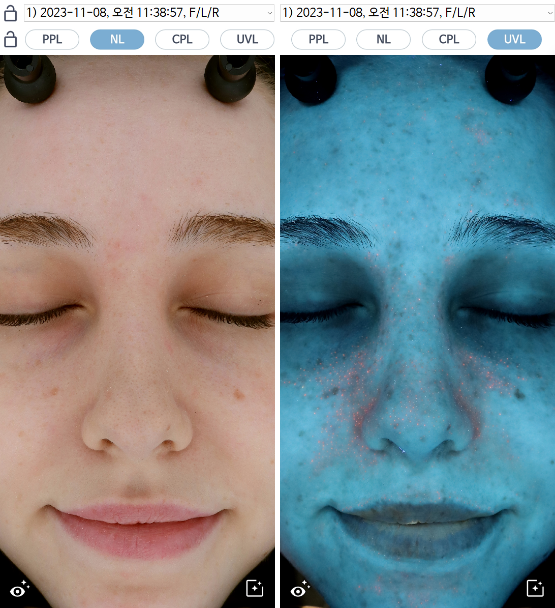 ▲ Differences in visible pigmentation under standard and UV light
385 nm: A UV Light Source for Observing Pigmentation and Porphyrins
JANUS Pro Hybrid and JANUS Pro Sunlike use a 385 nm UV light source.
385 nm is a long-wave UVA wavelength closer to visible light than 365 nm, with a lower relative UV photochemical hazard weighting under equivalent exposure conditions.
Porphyrins also strongly absorb light around 400 nm, so light sources near this wavelength range are used to observe porphyrin fluorescence.
JANUS Pro combines a 385 nm UV light source with its imaging system to support the analysis of UV pigmentation, sebum, and porphyrins.
This allows clinicians to focus consultations on pigmentation distribution while also reviewing sebum and porphyrin analysis results when needed.
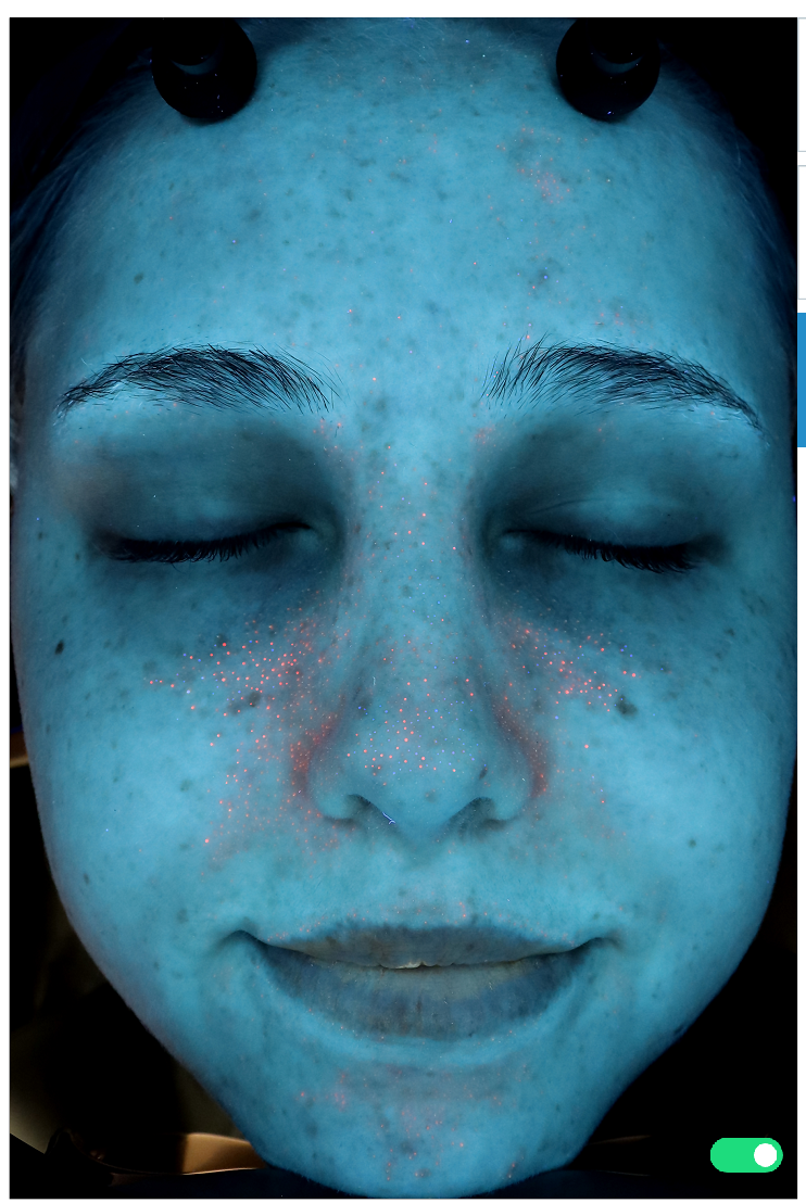 ▲ Image with detected porphyrins marked
Using the First Visit’s Images as a Reference for Future Consultations
Images taken at the first visit provide a reference for comparing subsequent skin changes. JANUS Pro can support the entire pigmentation consultation process, from showing the current skin condition to recording and explaining changes over time.
Presenting the skin’s appearance under standard light alongside pigmentation distribution observed in polarized and UV images helps patients understand their skin condition. At subsequent visits, imaging history allows clinicians to compare the same areas in earlier and current images and explain changes in pigmentation distribution and appearance.
Building a record of images taken under consistent conditions allows consultations to follow a consistent reference, from the initial assessment through follow-up reviews.