


Polarized Imaging in Skin Analysis: What’s the Difference Between Parallel and Cross Polarization Images?
Even when imaging the same skin, a skin analyzer can reveal different skin characteristics depending on how light is projected onto and captured from the skin.
Two representative imaging methods are parallel-polarized light (PPL) and cross-polarized light (CPL).
Simply put, PPL utilizes light reflected from the skin’s surface, making it useful for observing surface conditions. CPL reduces strong surface reflections, making it useful for observing skin color information, such as pigmentation and redness.
Parallel-Polarized Light (PPL) — Observing Surface Reflection and Texture
Observation depth: Skin surface
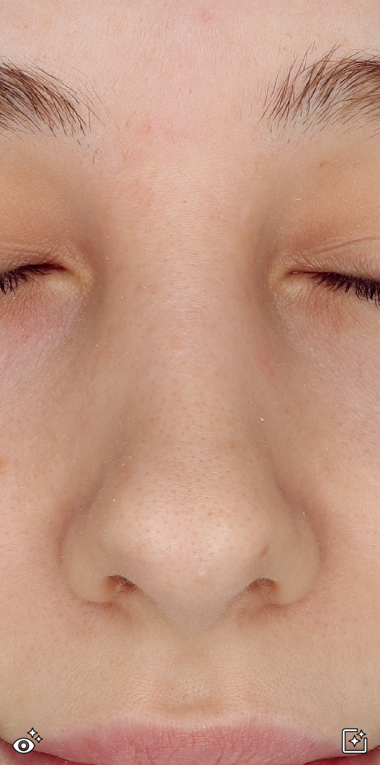
Parallel-polarized imaging is performed by aligning the polarizing filter in front of the light source with the polarizing filter in front of the camera in the same direction. With this method, a relatively large amount of light reflected from the skin’s surface remains visible in the image. It therefore helps reveal skin shine, reflections caused by sebum, fine surface irregularities, wrinkle contours, and skin texture.
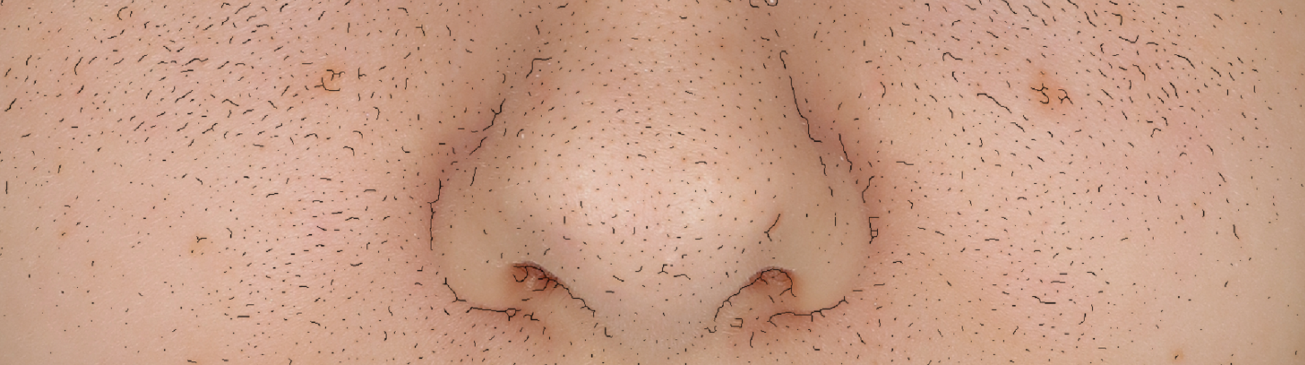
In simple terms, rather than looking beneath the skin, this imaging method helps determine how smooth or rough the skin surface appears and where light is strongly reflected.
It can be used to evaluate wrinkles and skin texture and to compare surface changes before and after cosmetic product use or treatment. However, surface reflections can also be affected by lighting, sebum, perspiration, cosmetics, and the imaging angle. Images should therefore always be captured under consistent conditions.
Cross-Polarized Light (CPL) — Observing Pigmentation and Redness
Observation depth (depending on skin tone): Approximately 0.1–0.8 mm (epidermis to upper dermis)
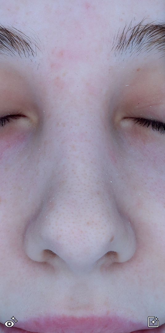
Cross-polarized imaging is performed by positioning the polarizing filter in front of the light source and the polarizing filter in front of the camera at right angles to each other.
When the two polarization axes are crossed, most of the strong light reflected directly from the skin’s surface is blocked. As a result, glare and shiny reflections are reduced, while color information from light scattered within the skin becomes relatively more visible.
In simple terms:
[Light → enters the skin → some is immediately reflected from the surface and blocked by the camera filter → some penetrates and scatters within the skin → exits the skin again → detected by the camera]
Because the polarization axes are crossed at 90° in CPL imaging, specular reflection directly from the surface is significantly suppressed, while scattered light whose polarization state has changed is detected more effectively.
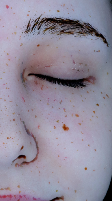
CPL is therefore useful for observing pigmentation, redness, and vascular red areas.
In darker skin tones or skin with higher pigmentation, overall image brightness and contrast may be reduced.
When Should PPL and CPL Be Used?
Neither method is inherently superior to the other; each serves a different observation purpose.
Imaging method / Main information observed / Simple explanation
PPL: Shine, sebum reflection, skin texture, wrinkles, and surface irregularities (emphasizes the skin surface)
CPL: Pigmentation, the depth and boundaries of pigmentation, redness, and vascular red areas (reduces surface reflection and emphasizes color differences)
PPL is useful for examining skin surface texture and reflection, while CPL is useful for observing pigmentation and red areas.
Therefore, using both types of images in a skin analyzer provides a more comprehensive understanding of the skin’s condition.